
Many women think of cancer the moment they hear the word 'tumour' during a breast ultrasound, but tumour is only a general term for a lump or mass and does not mean the same thing as cancer.
In fact, most tumours found in the breast are benign and do not invade the surrounding tissue, and the name breast cancer is used only when a tumour has been confirmed as malignant. Even so, the nature of a lump cannot be known from how it feels through the skin, which is why a detailed check with breast ultrasound is needed. This column sets out in turn what ultrasound can show, how benign tumours are observed, and when a biopsy becomes the next step.
01
Should cancer be suspected straight away when a lump is felt in the breast?
Key point
No. 'Tumour' is simply a general term for a lump or mass, and most of those found in the breast turn out to be benign. The name breast cancer is used only when a tumour has been confirmed as malignant, so it is important to understand exactly which diagnosis has been given.
Breast tumours are divided into benign and malignant, and most are benign growths that do not invade the surrounding tissue. Breast cancer is a diagnosis given only when a tumour has been confirmed as malignant, so it is worth knowing from the start that feeling a lump does not in itself mean cancer.
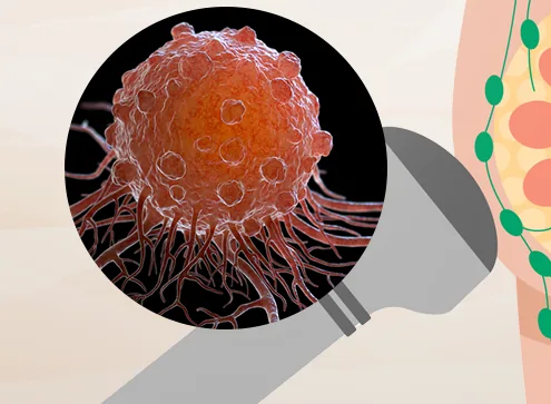
Touching the skin or looking at the breast gives no way of judging whether a tumour is benign or malignant, or what its size and shape are, so detailed imaging is needed. In a breast ultrasound, a probe sends out sound waves and the reflected waves are turned into an image, showing in real time the internal information that the hands cannot detect.
02
Can a breast ultrasound alone confirm whether it is breast cancer?
Key point
Ultrasound alone cannot confirm breast cancer. It can show in detail whether a lump is filled with fluid or closer to inflammation, and what its size and borders look like, but telling whether it is cancer requires moving on to a next step such as a biopsy.
A breast ultrasound shows in detail whether a tumour is filled with fluid or closer to inflammation, and what its size, borders and shape look like, which helps gauge how closely it may be related to cancer. This information is then used to decide whether to move on to the next step or simply to continue observation.
The findings considered more closely linked to malignancy can be summarised in a few points. The more an irregular or taller-than-wide shape, a rapid change in size over a short period and strong blood flow appear together, the more likely it is that further detailed testing will follow. Each finding is listed below.
- Shape — the border is not smooth and looks jagged or irregular
- Orientation — a lump that has grown taller than it is wide is examined more carefully
- Speed of change — the size increases rapidly within a short period
- Blood flow — strong blood flow around the tumour is seen at the same time
03
How is a benign tumour without pain managed, and how is the diagnosis confirmed?
Key point
For a benign lump that causes no pain, follow-up observation at intervals of several months to 2 years is usually enough on its own. If no clear change in size or shape appears as time passes, there is no particular need to rush into treatment straight away.
Even among benign tumours, a careful approach is needed when one suddenly grows to 2 cm or more, or when one side becomes elongated and the shape changes. It is also worth noting that a phyllodes tumour, which has a leaf-like pattern, involves fibrous and epithelial tissue growing together and is classified as benign, borderline or malignant.
Because ultrasound alone cannot establish the nature of a tumour, a core needle biopsy is needed. A long, thin needle is used to take a sufficient amount of tissue from the lesion, which gives high diagnostic accuracy and makes the test a key step in deciding the direction of treatment, so it deserves to be taken seriously. If treatment has been recommended straight away without a biopsy, comparing clinics against the criteria below can also help.
- Explanation of the diagnosis — whether the current condition and the diagnosis are explained accurately and in enough detail
- Timing of tests — whether the appropriate tests are carried out at the moment they are needed, without being missed
- Direction of treatment — whether an appropriate course of treatment is proposed and long-term follow-up is provided afterwards
Doctor's view
How the doctor decides
Seeun Cho, Medical Director · Obstetrics & Gynecology Specialist
I first establish whether it is benign or malignant
I often meet patients who are badly shaken by the single word 'tumour', and each time I tell them that the first step is to establish whether it is benign or malignant. I believe it is too early either to feel reassured or to worry on the basis of how a lump feels through the skin, so I do not judge by the presence or absence of pain alone. I look at the borders and shape seen on ultrasound together and then decide the next step.
I do not recommend treatment before a biopsy
I do not draw up a treatment plan before the diagnosis is confirmed. I believe that only after the nature of the tumour has been checked with a core needle biopsy can I judge whether removal is needed or observation is enough. When a patient has been advised elsewhere to have treatment without a biopsy, I sometimes suggest hearing another opinion.
I set the follow-up interval according to whether there is change
I do not tell patients to feel completely reassured just because there is no pain. I check for changes in size and shape at intervals of between several months and 2 years, and if a change is confirmed, I plan the next test again to match that point in time.
I believe care should continue after treatment
I do not think that care ends with removal or a biopsy. Because I believe a tumour needs to be watched over the long term with the possibility of recurrence in mind, I try to explain the diagnosis, the tests and the follow-up care thoroughly.
Summary
In summary
Hearing the word 'tumour' during a breast ultrasound is not a reason to worry about cancer straight away. Most tumours are confirmed as benign, and when there is no pain, follow-up observation at intervals of several months to 2 years is often enough. However, when a lump suddenly grows to 2 cm or more or changes shape, or when findings such as an irregular form or strong blood flow are seen together on ultrasound, a core needle biopsy is needed to confirm its nature accurately. If a treatment plan has been recommended without a confirmed diagnosis, hearing an additional opinion before deciding is one option. When a lump is felt in the breast, or there is pain unrelated to the menstrual cycle, the place to start is a breast ultrasound to check the nature of the tumour.
FAQ
Frequently asked questions
Does a tumour seen on breast ultrasound always mean surgery?
No. For a benign tumour with smooth borders and a uniform shape, follow-up observation at intervals of several months to 2 years is often enough, and the next step is reviewed again when a clear change in size or shape appears.
Can an ultrasound alone tell for certain whether it is breast cancer?
Ultrasound alone cannot confirm the diagnosis. It shows structural information such as size, shape and borders, which helps gauge how closely a lump may be related to cancer, and a next step such as a core needle biopsy is needed for confirmation.
What should be done if treatment is recommended without a biopsy?
Rather than deciding straight away, hearing an additional opinion at another medical institution is one option. With any tumour, it is difficult to decide with confidence whether treatment is needed without going through a biopsy.